Quezon Avenue Cor. Scout Magbanua St.
  Quezon City, Philippines, 1103
  (02) 372-3825 - 44
  [email protected]

Digital Infrared Thermal Imaging (D.I.T.I.) Section

A Non-invasive thermal biologic diagnostic test that assesses problems such as nerve irritation, soft tissue injuries, musculo-skeletal disorders and circulatory disorders which are often interrelated. Thermography does not show pain
because pain is a perception. It does, however show the thermal dysfunction that correlates closely with pain syndromes as well as normalization when the healing process takes place. It provides the clinician with helpful medical information in the differential diagnosis of complex pain syndromes, especially reflex sympathetic dystrophy (RSD).

It is based on the principle that all biologic systems emit heat energy in the form of of electromagnetic radiation, convection or conduction. It measures the body’s heat emission pattern by providing a heat map of the body that can be produced either by conduction or radiant energies reflecting cellular metabolism coming under micro euromuscular
control by pattern recognition. Thermography offers a scientific advancement for the clinician in detecting neuropathic, myofascial, circulatory, skeletal and psychogenic pain.

PRINCIPLE

At temperature above absolute zero ( -273 “C ) all bodies radiate infrared (heat) energy. Thermal energy is proportional to a molecular activity and metabolism. When focused on a sensitive detector shielded from ambient heat
by immersion in liquid nitrogen or other coolants, the electromagnetic IR ( Infrared ) signals from the body can be converted into electronic signals and processed to form a real time reproducible picture. This can be photographed
or videotaped for permanent patient record and comparative serial studies

CLINICAL BENEFITS OF THERMOGRAPHY:

  • Radiculopathy
  • Non-radicular muscle and fascicular disorder (muscle spasm, ligament and muscle tear or sprain, myofascial
  • syndrome)
  • Peripheral nerve injury
  • Hyperhidrosis
  • Entrapment neuropathy
  • Reflex sympathetic dystrophy (Sudeck’s Atrophy)
  • Inflammatory (arthritis, bursitis, rheumatism, synovitis, tendonitis)
  • Post-Operative (procedural follow-up)
  • Psychological (malingering, hysteria,
  • migraine)
  • Screening (pre-employment)
Magnetic Resonance Imaging (MRI) Section

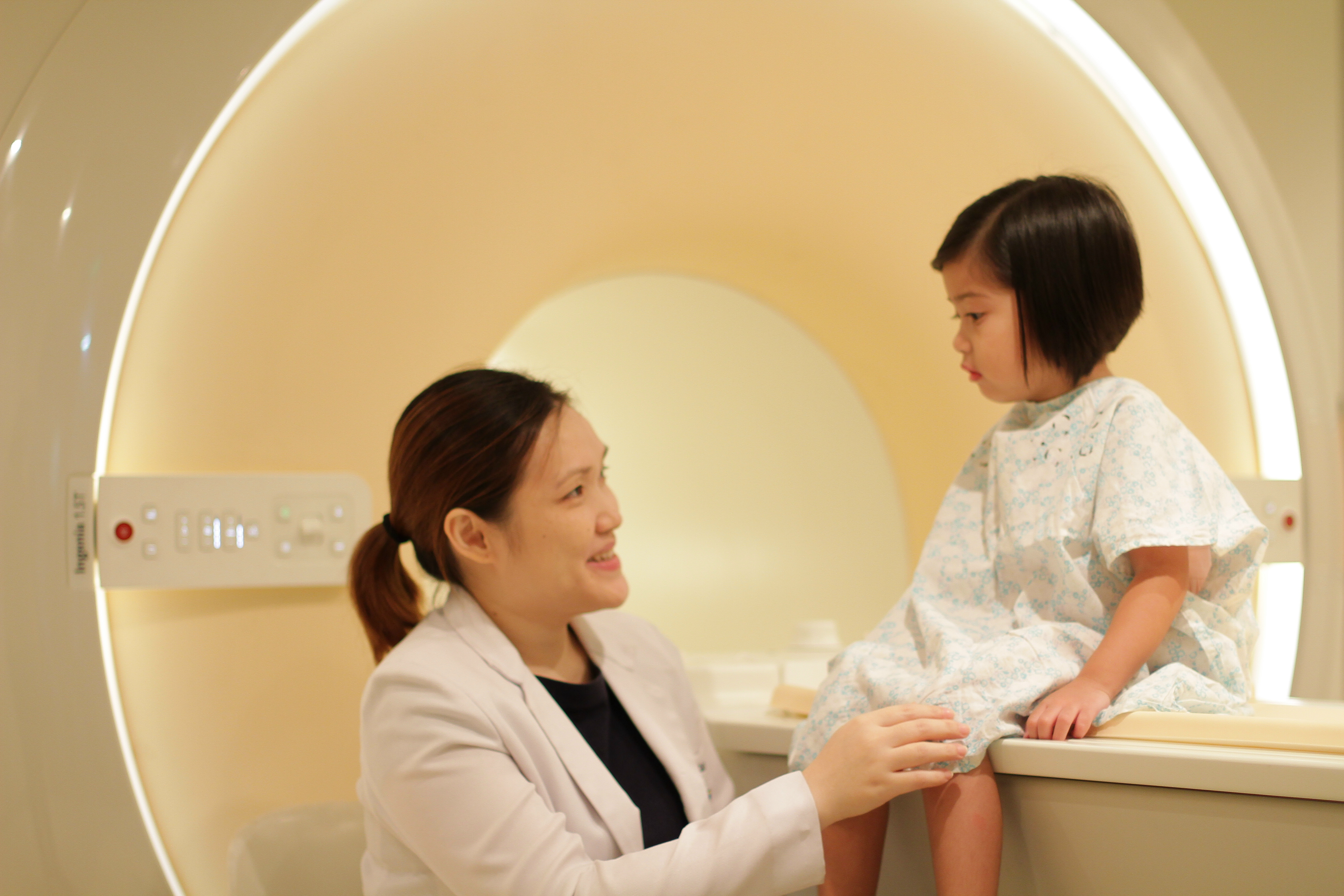
mriMRI or Magnetic Resonance Imaging is the latest and most modern non-invasive state–of-the-art diagnostic modality used by physicians to obtain vital information about a patient to guide them in their treatment. MRI utilizes magnetic fields, radio waves and computers to generate multi-planar images of the body.

With the new usage and applications continually being developed through on-going research, there have been many additional benefits of magnetic resonance imaging. The procedure is used for all parts of the body and is effective in the clinical evaluation of the following conditions:

  • Brain Disorders
  • Traumatic Injuries
  • Eye and Ear Abnormalities
  • Spine Diseases
  • Tumor Detection
  • Liver and other Abdominal Diseases
  • Injuries to the Extremities
  • Infection
  • Musculoskeletal Disorders
  • Blood Flow and Vessels Disorders
  • Breast Tumors

Capitol Medical Center’s Hitachi Aperto 0.4 Tesla Open MRI System complements the other diagnostic modalities already available in the hospital. The new open MR system has the highest magnetic field strength in its class. Being an “open” magnet, it does away with the anxiety associated with going into an enclosed space commonly experienced by patients with other MRI systems.

Examination time compared to other open MR units has been reduced to half, almost comparable with non-open MR system. The new MR also provides quality images matching those of higher field strength MR units.

FAQs

1. What is an Open-MR system ? What is the advantage of an Open-MR against a Closed – MR System?

An open-MR system uses a permanent magnet to generate the needed magnetic field thus, the magnetic field is low compared to the Closed-MR system. The new MR unit of Capitol Medical Center compared to all permanent magnets has a higher magnetic and gradient field strength allowing a shorter time for examination with superb image resolution. The open-system also answers the problem of claustrophobic patients. Nervous patients can be accompanied by a loved one or a family member during the examination.

2. What are the preparations needed before undergoing an MR examination?

There is no special preparation required before an MRI examination. The patient may eat normally and go about their daily routine. They may continue taking their medications as prescribed by their doctors.

3. Are there any contraindications in doing an MRI?

Physicians and our technical staff will screen patients for any surgically implanted metallic or electronic devices.

Pregnant women may undergo MRI examination if deemed necessary by their attending physician, since there are no documented harmful effects on the fetus.

4. Is there a need for intravenous contrast?

MRI examinations may be done with intravenous contrast depending on the information sought by the attending physician. The use of intravenous contrast allows clear visualization and delineation of abnormalities in many parts of the body. The intravenous contrast used in MRI is Gadolinium-based and is relatively safe among patients with renal dysfunction or previous allergic reaction to iodine-based contrast.

5. How long does an MR examination last?

On the average, each examination may last from 25 to 45 minutes depending on the request of the attending physician. Examination of multiple regions will take a much longer time.


CAPITOL MEDICAL CENTER, INC.
Quezon Avenue Cor. Scout Magbanua St.
Quezon City, Philippines, 1103
(632) 372-3825 - 44

CAPITOL MEDICAL CENTER
College of Nursing

No. 4 Sto. Domingo Avenue, Quezon City, Philippines 1100

Capitol Medical Center - College of Nursing

packages