Newborn Hearing Screening Program

The Newborn Hearing Screening Program (NHSP) of Capitol Medical Center under the Department of Pediatric aims to identify moderate, severe and profound hearing impairment in newborn babies. The program automatically offers all parents, the opportunity to have their baby’s hearing tested shortly after birth. Early identification, via the program, gives babies a better ‘life chance’ of developing speech and language skills and of making the most of social and emotional interaction for an early age.

Computed Tomography Scan

The Department of Radiology of Capitol Medical Center has a dedicated team of Radiologists who strive to achieve the best possible Computed Tomography imaging at the lowest exposure dose under the guidance of Dr. Jackson U. Dy, head of CMC CT scan section.

The CT scan is the world’s first adaptive scanner in maintaining the intricate details of the images. Stemming from our commitment in providing the best care now and into the future, CMC is proud to bring the best technology in the world.

The Capitol Medical Center (CMC) Computed Tomography Section uses the 128-slice Toshiba Aquilion CX, one of the latest generations in a line of CT scanners. The said machine can scan the whole body in seconds and provide incredibly 3d images of any organ.

The Toshiba Aquilion CT is design to boost productivity and expand the number of clinical applications possible. It also provides ultimate platform for treating a broader range of patients by performing a wider range of procedures. Hi-definition reconstruction algorithms providing 0.35-mm spatial resolution and 2-mm at 0.3% low-contrast resolution. With these in mind, CT scan is one of the most important methods of radiological diagnosis. It will help the hospital expand its service to more physicians and specialist.

The CMC Department of Radiology can provide CT examinations like cancer detection, pulmonary angiography or pulmonary embolism, cerebral angiography and prefusion for stroke evaluation and CT colonagraphy. It delivers non-superimposed, cross-sectional images of the body, which can allow smaller contrast differences than conventional x-ray images. This allow better visualization of specific differently structured soft-tissue regions, which could otherwise not be visualized satisfactorily.

Facilities

state of the art

Capitol Medical Center is committed to providing the best health care for its patients. Throughout its 45 year history, CMC has always acquired state-of-the-art hospital equipment to better serve its patients.

Capitol Medical Center offers world-class healthcare that costs substantially less than those in developed countries, using the same technology delivered by competent doctors.


SPECIALTY CLINIC

 Breast Care Center         CT Scan
 Cardiac Catheterization Laboratory Ear, Nose & Throat Center
 EECP Section EEG Section
 Emergency Room EMG section
 Endoscopy Section Eye Center
 Non-Invasive Laboratory /Heart Station  Kidney Center
 Laparoscopy Section Lunar Bone Densitometry
 Magnetic Resonance Imaging (MRI) Nuclear Medicine Section
 Nutrition Clinic OB Sonology
 Pathology Department Plastic & Cosmetic Surgery Center
 Pulmonary Section Radiology Department
 Rehabilitation Medicine Department Skin Center
 Sleep Center Ultrasound Section
 Women’s Care Center


RADIOLOGY DEPARTMENT      LABORATORY DEPARTMENT   
X-Ray           HIV Screening Test      
CT Scan (128 slice)         Blood Bank        
Magnetic Resonance Imaging (MRI) Open Type   Pathology Services      
Ultrasound             Anatomic Pathology    
Nuclear Medicine                Surgical / Cytology    
Bone Densitometry           Clinical Pathology      
Breast Care Center                Clinical Chemistry    
  Digital Mammography         Hematology and Blood Coagulation  
  Breast Ultrasound                Microbiology      
Gamma Camera                Immunology      
  Thyroid Scan                Parasitology      
  Cardiac Scan                Clinical Microscopy    
  Thallium                      
  Pulmonary Scan         EAR, NOSE & THROAT CENTER 
  Brain Scan           Hearing Accuracy Tests    
  Genito-Urinary Scan           Eustachian Tube Function    
  Gastro-Intestinal Scan         Eardrum Visualization    
  Hepatobiliary Scan                Tumor Detection      
  Bone Scan                Hearing Aid Dispensing    
  Scintimammography              Nasal Obstruction    
  In-Vitro Tests                Nose and Nasopharynx Visualization  
  Radio Fluoroscopy                     Rhionopharyngoscopy    
  Angiography                Larynx and Vocal Cords Visualization  
                          Laryngoscopy      
EYE CENTER                   Swallowing Assessment    
  Cataract Extraction                Voice Evaluation      
  Eye Refraction                     Videostroboscopy    
  Eye Diseases                Voice and Swallowing Disorders  
  Laser                       Rehabilitation      
                     Botox        
REHABILITATION DEPARTMENT               
  Physical Therapy         CARDIOVASCULAR CENTER
  Occupational Therapy       Catheterization Laboratory    
              Non-Invasive Laboratory    
PULMONARY LABORATORY       Holter Monitor      
  Chest Physio-Therapy         2D Echo Doppler      
  Ultrasonic Nebulization         Stress Test      
  PFT / IPPB           EKG        
  PEFR Determination         Peripheral Vascular    
  Body Plethysmograph                  
              DELIVERY ROOM  
OPERATING ROOM         High Risk        
  Laparoscopic           Cancer Clinic      
       Cholecystectomy           VIP Room      
  Endoscopy           Infertility / Menopause    
  SICCU             OB-Ultrasund      
  PACU                      
  Recovery Room         CHILD DEVELOPMENT UNIT  
                Speech Therapy      
NEUROPHYSIOLOGY LABORATORY     Occupational Therapy    
  Electromyography (EMG)                  
  Electroencephalographic (EEG)     NUTRITION CLINIC    
                         
EXECUTIVE CHECK-UP / WELLNESS PROGRAM KIDNEY TRANSPLANT CENTER
                         
THORACIC & CARDIOVASCULAR SURGERY KIDNEY / DIALYSIS CENTER
                         
ENHANCED EXTERNAL COUNTERPULSATION EMERGENCY ROOM (ER)
                         
NEONATAL INTENSIVE CARE UNIT (NICU) STROKE UNIT
                         
PEDIATRIC INTENSIVE CARE UNIT (PICU) SPINE CENTER    
                         
MEDICAL INTENSIVE CARE UNIT (MICU)  SKIN CENTER    
                         
OUT-PATIENT DEPARTMENT (OPD)    SLEEP CENTER     
                         
PHARMACY                    


Intensive Care Unit

icu

IMCU – MICU

6th Floor, CMC V Bldg.
372-3825 local 4613-4614

Ultrasonography (Ultrasound)

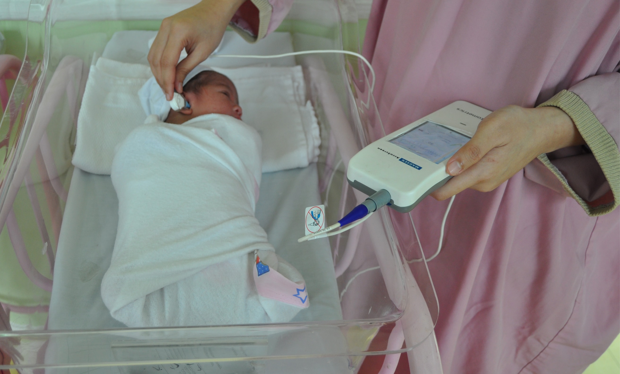
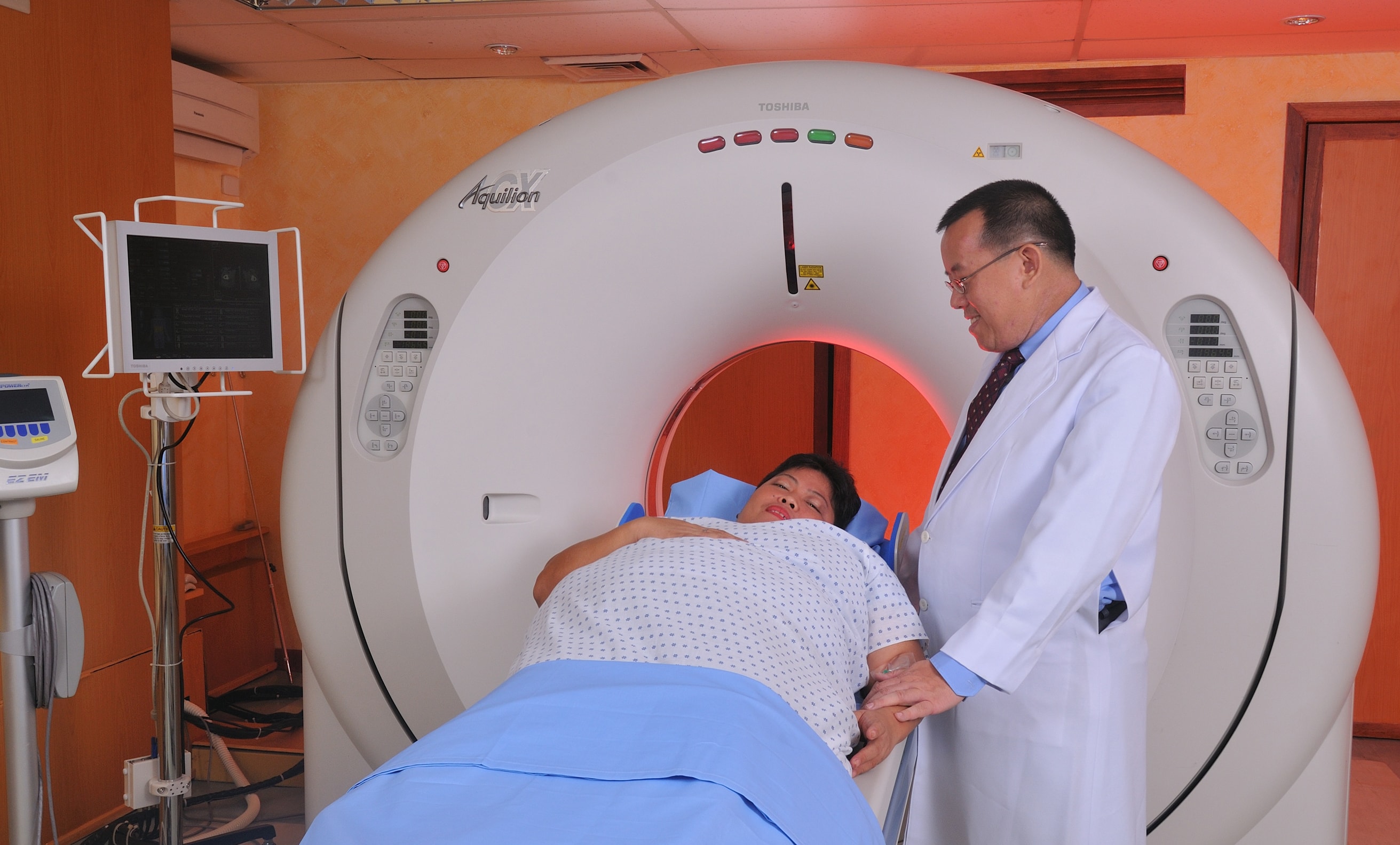
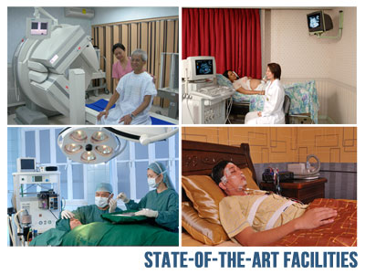
The section of Ultrasound is offering the whole range of diagnostic ultrasound examinations using state-of-the-art ultrasound machine that delivers outstanding diagnostic confidence. Virtually all parts of the body maybe examined using the systems, including neonatal brain, carotids, abdominal or peripheral blood vessels, thyroid glands, breast, prostate, testes, muscles, joints, soft tissues, chest abdomen (liver, gallbladder, bile ducts, pancreas, spleen, kidneys, appendix, etc.) and pelvis (uterus, ovaries and pregnancy).

Ultrasound-guided percutaneous biopsy of abdominal, thyroid or breast masses or tumors; needle aspiration or drainage of abscess, cyst, pleural effusion or and transrectal biopsy of the prostate are being performed with great precision and accuracy.

The service is manned by well-trained and experienced radiologists who are leaders in their field, and by qualified and competent ultrasonographers and other support staff.

For maximum benefit and accuracy, all examinations are carefully performed or personally supervised and checked by physician - radiologists.

No patient is allowed to leave the section without a proper ultrasound diagnosis or impression.

What is Ultrasonography ?

Ultrasonography is the use of high frequency sound waves to view organs and tissues inside the body. The pulses of sound waves are transmitted into the body using a transducer or probe. As they bounce off tissues or organs, the ultrasound machine captures the returning sound waves and creates a computerized picture of structures being examined.

Ultrasonography allows the evaluation of the size, shape, location, echo intensity (grays and blacks), and homogeneity of body structures. With this formation a list of possible causes of disease can be made and further diagnostic or therapeutic plans can be developed. Specific diagnosis may be determined; however, biopsy or needle aspirates are frequently required. Ultrasound can be very helpful guidance tool for biopsy or fine needle aspiration.


Subcategories